EMAT is one of the leading electron microscopy centers in the world and has a vast expertise in both fundamental and applied electron microscopy. EMAT has several state of the art electron microscopes including two aberration corrected, high end FEI-Titan instruments and a dual beam FIB.
EMAT provides access to a Helios NanoLab 650 to prepare TEM specimens with in-situ liftout. This instrument is a versatile, high-performance DualBeam system containing a Ga+ focused ion beam (FIB) together with a FEG extreme high-resolution scanning electron microscope (SEM) and Si-drift EDX detector for element analyses. In addition, the system is equipped with a Gas Injection System (GIS) containing a Pt source and an Omniprobe for micro-manipulation of the specimen. This device provides essential preparation for the majority of samples to be studied in installation 1.
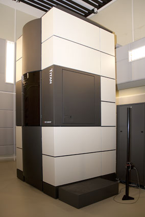
EMAT hosts two state-of-the-art FEI Titan3 microscopes that are both aberration corrected. The resolution in TEM is as low as 50 pm and in STEM 80 pm can be reached. The accelerating voltage is 300kV, but the instruments are also aligned to operate with atomic resolution at low voltage, which is crucial when investigating soft materials. Chemical analysis can be performed by EELS. Both instruments are equipped with a monochromator that lowers the energy spread of the electrons below 0.1 eV. As an alternative to EELS, a novel and highly efficient EDX system is available. The high efficiency of this EDX detector allows to record atomic resolution EELS and EDX data simultaneously. This unique feature is ideal to map the atomic positions of both heavy and light atoms. Special sample holders for electron tomography (double tilt rotation holder) as well as reconstruction software are available. Recently, two state-of-the-art direct electron detectors were installed. These detectors enable the acquisition of TEM images and EELS spectra using a drastically reduced electron dose. They also offer the potential for rapid read-out, enabling so-called 4D STEM imaging which vastly increases the amount of data that can be mined for content as compared to previous setups that typically resulted only in 2D images. This dramatically improves the information content per incoming electron dose which is of utmost importance in the imaging of beam sensitive materials. A set of liquid and gas environmental sample holders is being installed and will be available to the project, offering the unique combination of ultimate performance with realistic liquid and gas environments, providing a key benefit in e.g. catalysis research.
Scientists at EMAT are the experts in the field of energy filtered imaging (EFTEM) and of electron energy loss spectroscopy (EELS). The software package EELSMODEL is widely considered as the optimal approach for EELS quantification and has been developed at EMAT. This unique knowledge is made available to the ESTEEM3 users. Also concerning 3D imaging and quantification, EMAT is world leading. Different advanced treatments for the interpretation of electron tomography results have been developed at EMAT and will be available in the framework of TA. Last but not least, EMAT has an excellent theoretical group, responsible for supporting and validating the experimental results. The software packages StatSTEM and MULTEM, both developed at EMAT, will provide ESTEEM3-users with well-established methods to quantify unknown structure parameters from experimental images and to accurately simulate complex TEM calculations in a time efficient manner.